from rabbit, purified by affinity chromatography
NA.41
primary antibodies
human
rabbit
polyclonal
western blot: suitable
human ... BIRC7(79444)
Q96CA5
Affinity purified
Anti-Birc7/LIVIN detects levels of Birc7/LIVIN proteins & has been published & validated for use in WB.
Unless otherwise stated in our catalog or other company documentation accompanying the product(s), our products are intended for research use only and are not to be used for any other purpose, which includes but is not limited to, unauthorized commercial uses, in vitro diagnostic uses, ex vivo or in vivo therapeutic uses or any type of consumption or application to humans or animals.
Concentration: Please refer to the Certificate of Analysis for the lot-specific concentration.
Livin, also known as “baculoviral IAP repeat-containing protein 7” (Birc7) and \KIAP,\ is a member of the IAP (inhibitors of apoptosis) family of proteins characterized by highly conserved baculoviral IAP repeats (BIR). In addition, Livin contains one RING-type zinc finger domain. Livin has been detected in the heart, placental, lung, lymph node, ovary, spleen, and in various types of solid tumors. It may suppress apoptosis induced by TNF, viral infection, drugs, and growth factor withdrawal, by inhibiting caspase activity (caspase 3, 7, and 9) and activating a MAPK/JUN pathway. Paradoxically, Livin may also function as a pro-apoptic factor in a truncated form (tLivin) that is cleaved by caspase 3 from the full-length protein. The pro-apoptotic effects of Livin may depend on its RING domain. Livin may play a role in non-Hodgkin lymphoma.
Affinity purified
KLH-conjugated linear peptide corresponding to the BIR repeat region of human Birc7/LIVIN.
Control Human spleen tissue lysate
This antibody recognizes the BIR repeat region of Birc7/LIVIN.
Stable for 1 year at 2-8°C from date of receipt.
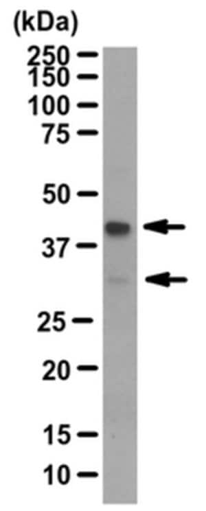
Evaluated by Western Blot in human spleen tissue lysate.Western Blot Analysis: 1 µg/mL of this antibody detected Birc7/LIVIN in 10 µg of human spleen tissue lysate.
~39 kDa, ~32 kDa observed. Two isoforms at 32 kDa and 30 kDa are known to exist. This protein has been observed at ~39 kDa (alpha) and ~37 kDa (beta) due to post translational modification (Nachmias, B., et al. (2007). Apoptosis. 12(7):1129-1142.).

 收 藏
收 藏